Brain Fog — Causes & Treatment
Brain Fog Is Not in Your Head. It Is in Your Cells.
If your thinking has slowed, your words go missing, and you feel mentally underwater — your brain is running low on cellular energy. Here is what is actually happening, and how we address it.
Virginia Beach • Blacksburg • Telemedicine Across Virginia

The Problem
Brain Fog Is a Symptom. Your Doctor Is Treating It Like a Diagnosis.
You have probably been told your labs are normal. Your thyroid is "fine." Your hormones are "within range." And yet you cannot finish a sentence, you lose words mid-thought, and the mental clarity you used to take for granted is gone.
This is brain fog. And it is not a personality trait, a mood disorder, or the inevitable cost of getting older. It is a measurable failure of cellular energy production inside your brain.
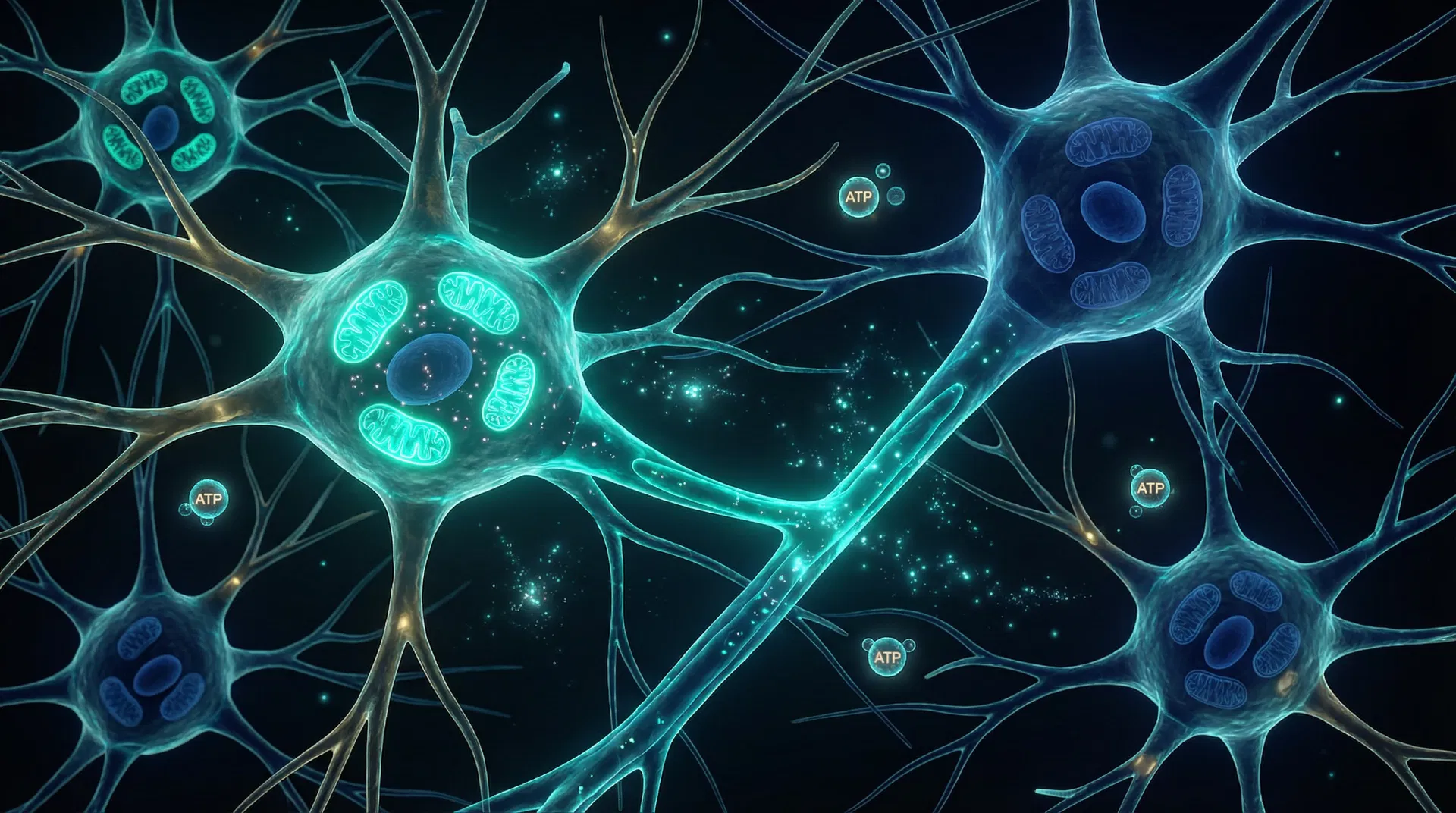
The brain is the most metabolically demanding organ in the body. It accounts for roughly 20 percent of your total energy consumption despite representing only two percent of your body weight. That energy comes from one source: ATP, produced inside your mitochondria. When mitochondrial output drops, brain function is the first place you feel it.
The cognitive symptoms — slow processing, word retrieval failure, difficulty concentrating, mental fatigue after simple tasks — are not vague or subjective. They are predictable downstream effects of impaired cellular energy metabolism. Once you understand the mechanism, the path forward becomes clear.
The Mechanism
The Cellular Energy Explanation for Brain Fog
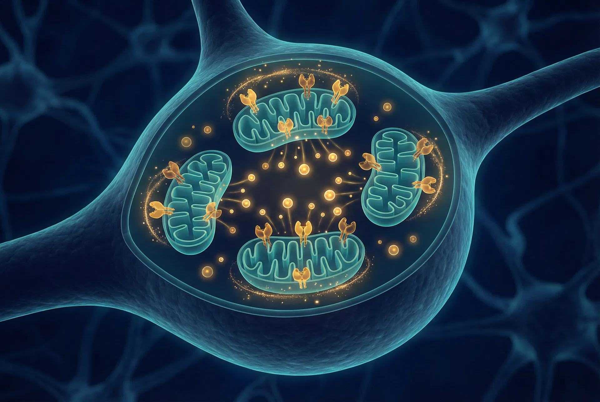
Your neurons depend on a continuous, high-volume supply of ATP. Unlike muscle cells, neurons cannot rest and recover between demands. They operate around the clock — managing neurotransmitter synthesis, maintaining electrical gradients across cell membranes, supporting axonal transport, and running the constant background processing that constitutes cognition.
When cellular energy production becomes insufficient to meet those demands, the brain activates a protective response called the Cell Danger Response (CDR). The CDR is a conserved biological survival program: when cells detect insufficient resources or threat, they downregulate normal metabolic activity and shift into a defensive posture.
In the brain, CDR activation means reduced synaptic signaling, impaired neurotransmitter recycling, slower neural conduction, and suppressed neuroplasticity. This is not damage. It is a deliberate downshift to protect the cell. But from the outside, it feels exactly like brain fog.
The Drivers
What Disrupts Mitochondrial Output in the Brain
Mitochondrial Nutrient Deficiencies
B vitamins (especially B1, B2, B3, B5, B12), CoQ10, magnesium, carnitine, and alpha-lipoic acid are required cofactors at every stage of the electron transport chain. Deficiencies bottleneck ATP output.
Neuroinflammation
Chronic low-grade inflammation elevates microglial activation, impairs mitochondrial membrane integrity, and competes with neurons for metabolic resources.
Oxidative Stress
The brain is particularly vulnerable to reactive oxygen species because of its high fat content and high oxygen consumption. Mitochondrial oxidative damage directly impairs electron transport efficiency.
Impaired Glucose Regulation
The brain runs on glucose. Insulin resistance, blood sugar dysregulation, and impaired glucose transport across the blood-brain barrier all reduce the fuel supply available for ATP synthesis.
Hormonal Decline
Estradiol and thyroid hormone are direct regulators of mitochondrial function in neurons. Their deficiency is not a peripheral concern. It is central to brain energy failure.
These factors do not operate in isolation. In most of the patients I see, several of them are present simultaneously. The cumulative effect on brain energy output is significant — and almost always missed by conventional screening because each individual marker sits just inside the "normal" range.
Why Hormones Matter
Why Hormones Matter for Brain Fog
Hormones are not a separate conversation from cellular energy. They are part of the same conversation. Estradiol and thyroid hormone are among the most powerful regulators of mitochondrial function in the human brain. When either is insufficient, neuronal energy production suffers directly.
Estradiol: The Brain's Primary Metabolic Hormone
Estradiol is not a reproductive hormone that happens to affect the brain. It is a neurological hormone with reproductive functions. The distinction matters because it explains why cognitive symptoms emerge so early in perimenopause — often years before the final menstrual period — and why conventional medicine consistently underestimates the impact of estradiol decline on brain function.
Estrogen receptors — specifically ERα and ERβ — are distributed throughout the brain in regions directly governing cognition, memory, and executive function: the prefrontal cortex (executive function, working memory), the hippocampus (memory consolidation), the amygdala (emotional regulation), and the cerebral cortex (processing speed, language).
But estrogen receptors are not only on neurons. They are on the mitochondria themselves. Both ERα and ERβ are expressed on the inner mitochondrial membrane. This means estradiol acts directly at the site of energy production inside brain cells.
Mitochondrial Biogenesis
Upregulates PGC-1α, the master regulator of new mitochondria production.
ETC Efficiency
Upregulates Complex I and Complex IV activity for more efficient ATP synthesis.
Anti-Neuroinflammation
Suppresses microglial activation. Declining estradiol increases neuroinflammation.
Acetylcholine Synthesis
Upregulates choline acetyltransferase — the enzyme producing the neurotransmitter of memory.
BDNF Regulation
Supports neuroplasticity and synaptic strength via brain-derived neurotrophic factor.
Myelin Integrity
Maintains myelin sheaths around axons. Degraded myelin slows neural conduction velocity.
The perimenopausal brain fog that patients describe — the word retrieval failure, the cognitive slowness that feels out of character, the sense of "not being sharp anymore" — maps directly onto this mechanism. The brain is running on a reduced energy budget because the hormone that powered its mitochondria is declining. This is not psychiatric. It is metabolic.
This is also why the timing matters. The critical window for neuroprotection is early perimenopause — before sustained neuroinflammation and mitochondrial decline become entrenched. Intervention is more effective, and protective effect is more durable, when addressed before the system has been in deficit for years.
Testosterone and Male Brain Fog: The Dual-Mechanism Problem
Brain fog is not a women's health issue. Men experiencing andropause — the gradual decline of testosterone that begins in the early forties and accelerates through the fifties and sixties — report the same cognitive symptoms: word retrieval failure, slowed processing speed, difficulty sustaining concentration, and a persistent sense of mental cloudiness that was never there before.
The incidence of testosterone deficiency is approximately 20 percent in men aged 60 and increases to 50 percent by age 80. Among men over 45, nearly 39 percent have total testosterone below 300 ng/dL — the clinical threshold for hypogonadism. And low testosterone is independently associated with a 48 percent increased risk of Alzheimer's disease.
What makes this mechanism particularly important is that testosterone does not affect the brain through a single pathway. It operates through two distinct, parallel mechanisms — both of which must be understood to address male cognitive decline effectively.

Pathway 1
Direct Androgen Receptor Action
Testosterone acts directly on androgen receptors (AR) in the brain — independent of estradiol. Research published in the Journal of Neurochemistry demonstrated that "androgens induce neuroprotection directly through the androgen receptor," confirming this is not solely an estrogen-mediated effect.
Through this pathway, testosterone directly increases ATP production in neuronal mitochondria, enhances antioxidant defenses to regulate redox homeostasis, improves synaptic plasticity in the hippocampus, maintains blood-brain barrier integrity, and supports cerebral vascular remodeling. DHT — a non-aromatizable testosterone metabolite — independently modifies neural circuits by altering excitatory spine synapses, particularly influencing spatial memory and cognitive processing.
Pathway 2
Aromatization to Estradiol in the Brain
The brain contains the enzyme aromatase, which converts testosterone to estradiol locally within neural tissue. This brain-derived estradiol then acts on the same ERα and ERβ receptors on mitochondrial membranes described in the estradiol section above — the identical mechanism that drives perimenopausal brain fog in women.
Research published in Neurology showed that "improvement in verbal memory induced by testosterone administration depends on aromatization of testosterone to estradiol." A landmark study found that testosterone improved working memory in aged subjects, but DHT (which cannot convert to estradiol) did not — confirming that working memory specifically requires this conversion pathway.
Different Cognitive Domains, Different Pathways
Research by Cherrier et al. revealed a critical distinction: aromatization of testosterone to estradiol regulates verbal memory in men, while non-aromatizable androgens regulate spatial memory. This means the brain fog symptoms men describe — word-finding difficulty, mental cloudiness, slowed processing — likely involve both mechanisms simultaneously.
This is why testosterone replacement in men can improve cognition through a dual mechanism: restoring direct androgen receptor-mediated mitochondrial support and restoring the brain's local supply of estradiol. It is not one or the other. It is both.
At The Johnson Center, we evaluate male brain fog through this dual-mechanism lens. We measure total and free testosterone, DHT, SHBG, and estradiol — because understanding both the androgen and the aromatization pathways is essential to identifying where the cellular energy deficit originates and how to correct it.
Thyroid: The Cell's Master Metabolic Regulator
Every cell in the body has thyroid hormone receptors. Neurons are no exception. Thyroid hormone — specifically T3, the active form — enters the cell nucleus and directly regulates the genes responsible for mitochondrial transcription and energy metabolism. Without adequate T3, mitochondria cannot produce ATP at the rate the brain requires.
The cognitive symptoms of thyroid insufficiency are among the most common, and most commonly missed, presentations in functional practice: slowed processing speed, difficulty with word recall, sustained mental fatigue after cognitive effort, difficulty concentrating, and depression that does not respond to antidepressants.
These symptoms emerge even in subclinical hypothyroidism — a TSH that sits in the upper range of normal, with free T4 and free T3 that are technically within reference range but functionally low. Most conventional thyroid screening stops at TSH. That is not sufficient to evaluate brain performance.
The conversion of T4 to active T3 is a separate, rate-limiting step that happens in peripheral tissues — including the brain itself. This conversion depends on selenium, zinc, iron, and adequate iodine. It is impaired by chronic stress, gut inflammation, and nutrient depletion. A patient can have a normal TSH and a T4 in the mid-range and still be functionally hypothyroid at the cellular level.
At The Johnson Center for Functional Health & Longevity, we do not screen thyroid function with TSH alone. We run a full panel: TSH, free T4, free T3, reverse T3, TPO antibodies, and thyroglobulin antibodies. We are looking at how well thyroid hormone is being produced, converted, and utilized at the cellular level — not whether a number clears an insurance threshold.
The Patterns
What Is Actually Driving Your Brain Fog
Brain fog is not a single-cause problem. It is the clinical expression of several converging cellular deficits. The work is to identify which ones are present in your specific biology and address them in the correct order.
Mitochondrial nutrient insufficiency
B vitamins, CoQ10, magnesium, carnitine. Often present without obvious dietary deficiency because demand is high and absorption is impaired.
Subclinical hypothyroidism with impaired T4-to-T3 conversion
Common in women over 40 with normal TSH and borderline free T3.
Perimenopausal estradiol decline
Presenting with cognitive symptoms months to years before menstrual irregularity begins.
Testosterone decline and andropause in men
Low testosterone impairs cognition through two pathways: direct androgen receptor neuroprotection and reduced brain-derived estradiol via aromatase conversion.
HPA axis dysregulation
Chronic cortisol elevation suppresses T3 conversion, impairs hippocampal function, and increases neuroinflammation simultaneously.
Intestinal permeability and systemic inflammation
Gut-derived LPS crosses into circulation and activates microglial cells in the brain. Neuroinflammation is often a gut problem in disguise.
Metabolic dysfunction and insulin resistance
Impaired glucose utilization in the brain is now recognized as a core feature of cognitive decline. It is detectable, measurable, and reversible.
None of these are mutually exclusive. Perimenopausal estradiol decline or andropausal testosterone decline, for example, increases neuroinflammation and reduces mitochondrial output — which then compounds the effects of any pre-existing nutrient insufficiency or thyroid dysfunction. The system failures amplify each other.
How We Test
How We Assess Brain Fog at The Johnson Center
Standard bloodwork does not provide enough information to understand a cellular energy problem. We use a layered diagnostic approach that maps what is actually happening at the metabolic level.
| Panel | What It Reveals |
|---|---|
| Organic Acid Test (OAT) | The most direct window into mitochondrial function. Measures metabolic byproducts of the Krebs cycle and electron transport chain. |
| Full Thyroid Panel | TSH, free T4, free T3, reverse T3, TPO antibodies, thyroglobulin antibodies. Not TSH alone. |
| Comprehensive Hormone Panel | Estradiol, progesterone, testosterone, DHEA-S, SHBG. Timed to cycle phase where applicable. |
| Adrenal & Cortisol Mapping | Four-point salivary cortisol to evaluate HPA axis rhythm and output. |
| Inflammation & Nutrient Panel | hs-CRP, homocysteine, ferritin, vitamin D, B12, zinc, magnesium. |
| GI & Permeability Testing | Neuroinflammation often originates in the gut. If the gut barrier is compromised, neurological inflammation persists. |

The results from these panels are interpreted together, not in isolation. A slightly low free T3 and a borderline ferritin and a cortisol curve that peaks too late — individually, these might each be dismissed. Together, they constitute a clear picture of why the brain is energy-insufficient.
The Protocol
Restoring Brain Function: The Cellular Intelligence Protocol™
The Cellular Intelligence Protocol™ (CIP) is the clinical framework we apply at The Johnson Center for Functional Health & Longevity. It addresses brain fog — and all cellular energy disorders — through three coordinated pillars.
Pillar 01
Cell Danger Response Resolution
Before the cell can return to full metabolic output, the signal driving the CDR must be resolved. This means addressing the upstream triggers — nutrient insufficiency, inflammation, hormonal deficiency, gut dysfunction — that told the cell it was under threat. This is not suppression. It is removal of the cause.
Pillar 02
Bioenergetic Core Restoration
Once CDR signals are resolving, we rebuild mitochondrial capacity directly. This includes targeted repletion of mitochondrial cofactors (B-complex, CoQ10, carnitine, magnesium, NAD+ precursors), support for electron transport chain function, and — where indicated — thyroid optimization and estradiol restoration.
Pillar 03
PNI Integration
The psychoneuroimmunology layer addresses the bidirectional relationship between the central nervous system, the immune system, and the endocrine system. Chronic psychological stress is a direct input into both HPA axis dysregulation and neuroinflammation. This pillar runs parallel from the first visit.
Brain fog resolution follows a characteristic sequence. Most patients notice improved sleep quality and reduced mental fatigue first — typically within the first four to eight weeks. Sustained cognitive improvements in processing speed, word retrieval, and concentration follow as mitochondrial capacity rebuilds. The timeline is individual. The mechanism is not.
What to Expect
It Is Not Fast. It Is Complete.
This is not a fast process. Mitochondrial recovery, hormonal optimization, and neuroinflammation resolution each operate on their own timeline. What we can tell you is that each of these systems is responsive to the right inputs — and that the brain is considerably more plastic and recoverable than conventional medicine typically represents.
The patients who recover most completely are those who commit to the full diagnostic and therapeutic protocol rather than treating individual symptoms. Brain fog is a systems problem. Addressing it with a single supplement or a thyroid prescription in isolation produces partial results at best.

Ready to Find Out What Is Driving
Your Brain Fog?
A single consultation provides more diagnostic clarity about your brain function than most people have accumulated in years of conventional care. We will review your history, interpret your existing labs, and determine exactly which cellular systems require assessment.
We work with patients in Virginia Beach, Blacksburg, and via telemedicine across Virginia.
Book a ConsultationFrequently Asked Questions
Common Questions About Brain Fog
Brain fog is a clinical term for a cluster of cognitive symptoms — impaired processing speed, difficulty with word retrieval, reduced working memory, poor concentration, and mental fatigue. It is not a diagnosis. It is a symptom of an underlying cellular energy deficit, usually involving mitochondrial dysfunction, hormonal insufficiency, neuroinflammation, or a combination of these.
Yes, and the mechanism is direct. Estrogen receptors are present on the mitochondria inside neurons. Estradiol regulates mitochondrial biogenesis, supports electron transport chain efficiency, suppresses neuroinflammation, and drives the production of acetylcholine — the neurotransmitter of memory and attention. When estradiol declines in perimenopause, brain energy production declines with it. The cognitive symptoms — word loss, processing slowness, difficulty concentrating — are the predictable result.
Thyroid hormone is a direct regulator of mitochondrial function in every cell, including neurons. Insufficient T3 reduces the brain's capacity to produce ATP. The cognitive symptoms of thyroid insufficiency — slow thinking, word retrieval difficulty, poor concentration, mental fatigue — are identical to other forms of brain fog. And subclinical hypothyroidism, where TSH is technically normal but free T3 is low, is common and commonly missed. We do not assess thyroid function with TSH alone.
We use an organic acid test to directly evaluate mitochondrial metabolic function, a full thyroid panel (not TSH alone), a comprehensive hormone panel, a four-point cortisol assessment, inflammation and nutrient markers, and intestinal permeability testing. These panels together provide a complete picture of the cellular systems involved.
Conventional medicine typically screens for serious pathology (thyroid disease, depression, anemia) and, finding nothing diagnostic, offers symptomatic treatment or reassurance. Functional medicine asks why the brain is energy-insufficient and addresses the cellular mechanisms responsible. The diagnostic depth is different. The therapeutic targets are different. The outcomes reflect that.
Early improvements — better sleep, reduced mental fatigue — typically occur within four to eight weeks of addressing the primary cellular drivers. Sustained cognitive recovery, including processing speed and memory, follows as mitochondrial capacity rebuilds. The timeline depends on how long the deficits have been present and how many systems are involved. It is not fast. It is complete.
Dr. Barbara Johnson
The Johnson Center for Functional Health & Longevity
Virginia Beach • Blacksburg • Telemedicine